Radiographs
We use radiographs or X-rays very regularly to gain more insight into what is going on in our little patient’s bodies. This form of diagnostic imaging allows us to assess the bones as well as look at the internal organs. What we can see will vary based on the type of animal and the positioning of the radiographs, as well as the disease process going. Radiographs are particularly useful for assessing bone structure, and to look for any broken bones or dislocations. We can often tell a bone is broken just with our physical examination, but a radiograph allows us to assess the break, how severely the bone is broken, where the break is, are there any other cracks or fissures in the bone – all of which will guide us in what treatment options will be available for the patient. Some broken bones can have a cast placed to help them heal, others require surgical pinning.
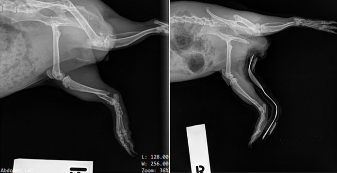
Here are some radiographs of a young guinea pig who broke her right hind leg. You can see the broken tibia in the photo on the left. A splint and bandage was applied and the radiograph on the right was to check if it was adequately holding the fracture in alignment – you can see it is doing a fantastic job of holding the bone pieces together to allow them to heal.
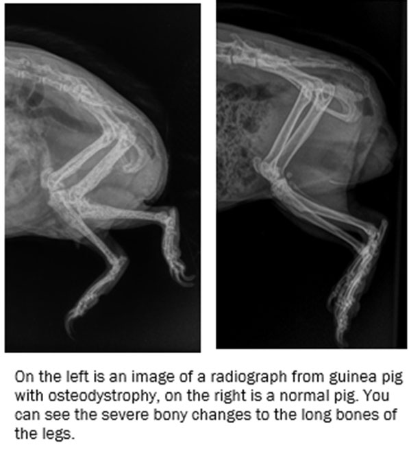
Radiographs allow us to assess the bones of our patients. Here you can see a guinea pig with osteodystrophy vs a normal guinea pig patient.
(look at the images below).
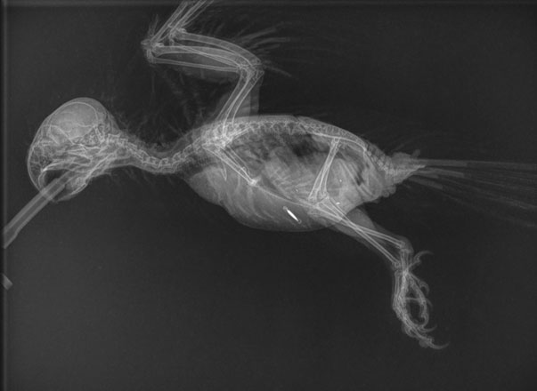
This radiograph is of a bird who has swallowed some metal pieces – you can see them in her caudal gut. You can also see the microchip that has been implanted in her pectoral muscles.
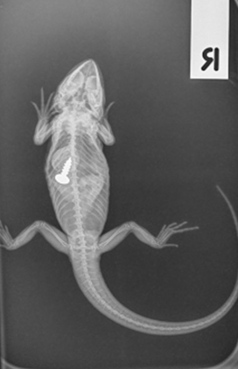
This is a radiograph of a bearded dragon that has eaten something she shouldn’t have!
The way we take radiographs will vary from species to species. Often to get the best images we need to anaesthetise our patients so that we can position them correctly. When we take radiographs of birds we need to stretch their wings away from the body – something that isn’t easily achievable without significant stress to the patient unless they are anaesthetised! In some circumstances we can do radiographs conscious or just use sedation, but it will vary from case to case. The veterinarian will discuss with you what will be the best option for your pet at the time of the procedure.
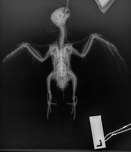
This is a radiograph of a cockatiel – can you spot what is abnormal? HINT – look at the right leg
Ultrasound
Ultrasound uses sound waves via a transducer to allow us to see internal organs. It is a great way to assess the soft tissues in the abdomen or coelom, as well as the heart. We can often do an ultrasound exam on a conscious patient although sometimes sedation or anaesthesia is required.
We most commonly use ultrasound to assess the body cavity. We can look for things such as:
- Fluid accumulation
- Abnormal masses
- Assess the urinary and reproductive tract
- Assess the heart
- Assessing for pregnancy and for foetal viability (pending foetal size)
Ultrasound is particularly useful to assess the reproductive status of female bearded dragons. By assessing the follicles growing on the ovaries we can see if they become static (follicular stasis) – a serious and sometimes life-threatening health issue.
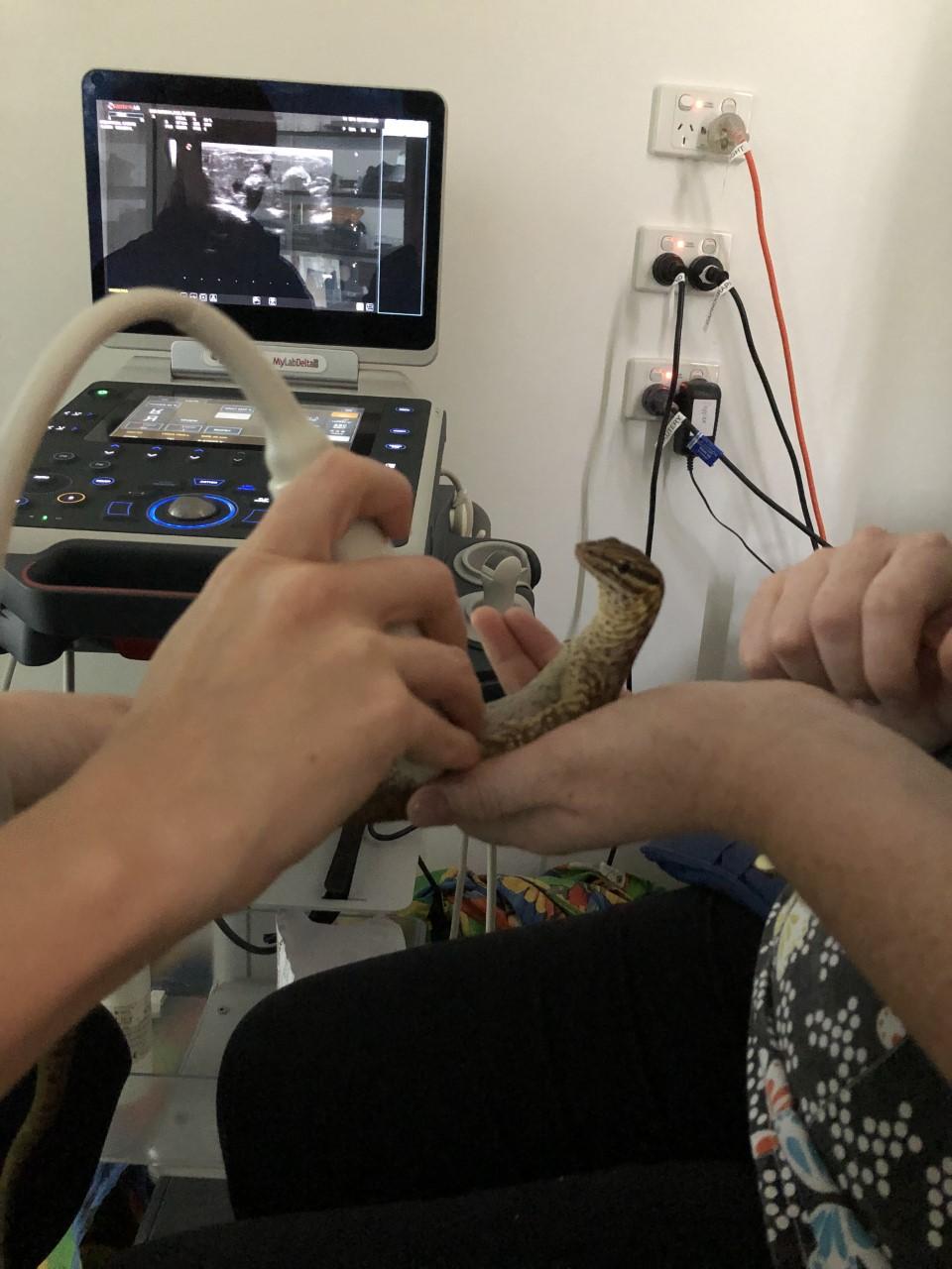
This little monitor lizard is having an ultrasound of her tummy
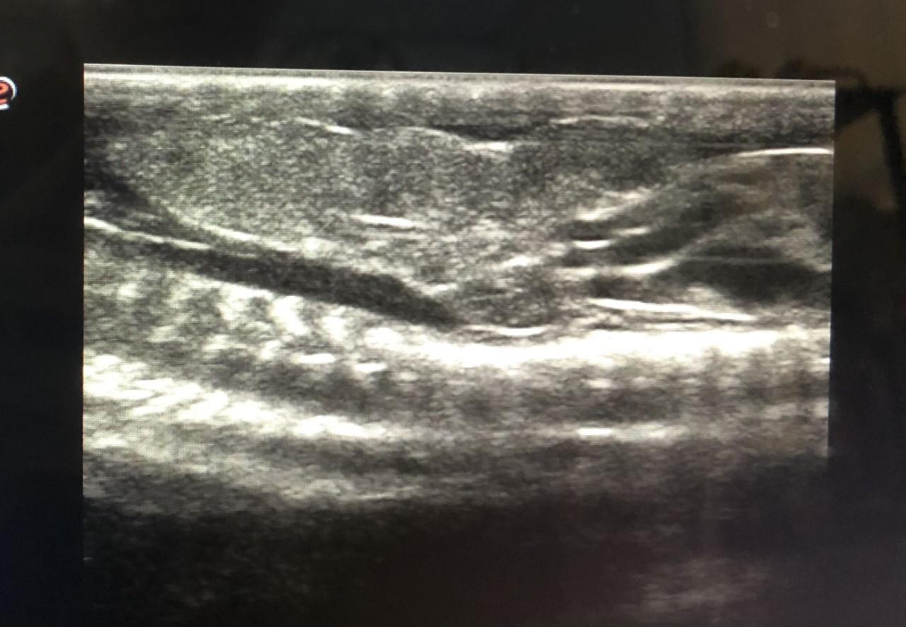
This ultrasound image shows a soft tissue mass dorsally with a large blood vessel running beneath it. It is useful to assess lumps like this prior to surgery to determine the best approach
CT Computed Technology
This type of diagnostic imaging uses a special machine that uses X-rays to create a 3D picture. We sometimes will do these after we have radiographs or ultrasound to give us further information on a patient. Although we don’t have a CT machine ourselves, we are able to take our patients offsite to have this done. CT allows us to have a 3D view of a patient and therefore can give more information on what is going on. It is particularly useful for looking at areas of the body that are ‘busy’ or have a lot of structures overlying each other, superimposed. Such as the skull and the body cavity. CT’s allow us to get a better view of what is going on and help us to assess prognosis and plan treatments
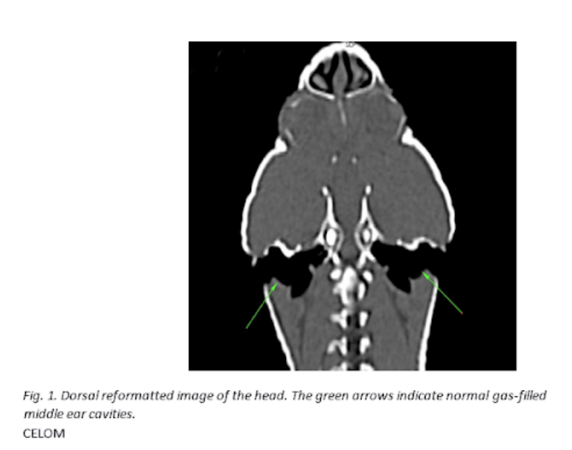
This is a CT scan image of a blue tongued skink
Endoscopy
Endoscopy is a form of imaging that uses a very small camera to physically look inside a patient! It requires a patient to be under general anaesthesia so they are very still. The very small camera can then be used to assess the patient. Regular uses include putting the camera down a patient’s trachea (or windpipe) to assess for any foreign bodies or lesions, or placing it inside a body cavity (such as the coelom) to allow visualisation of the organs and biopsy collection.
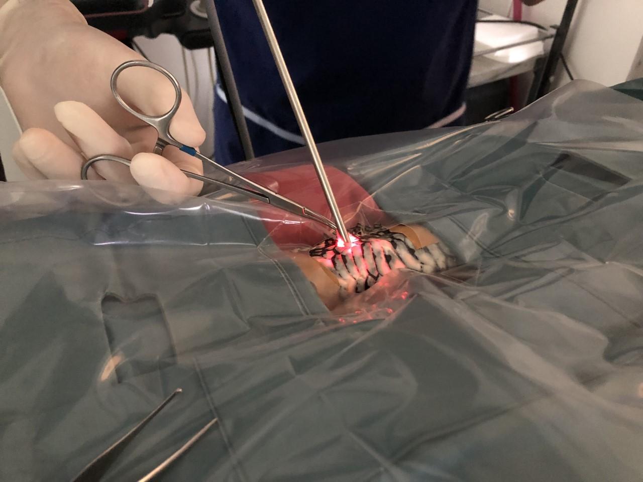
This is the endoscopy camera being inserted into the body cavity (coelom) of a python. In this case to assess the liver.
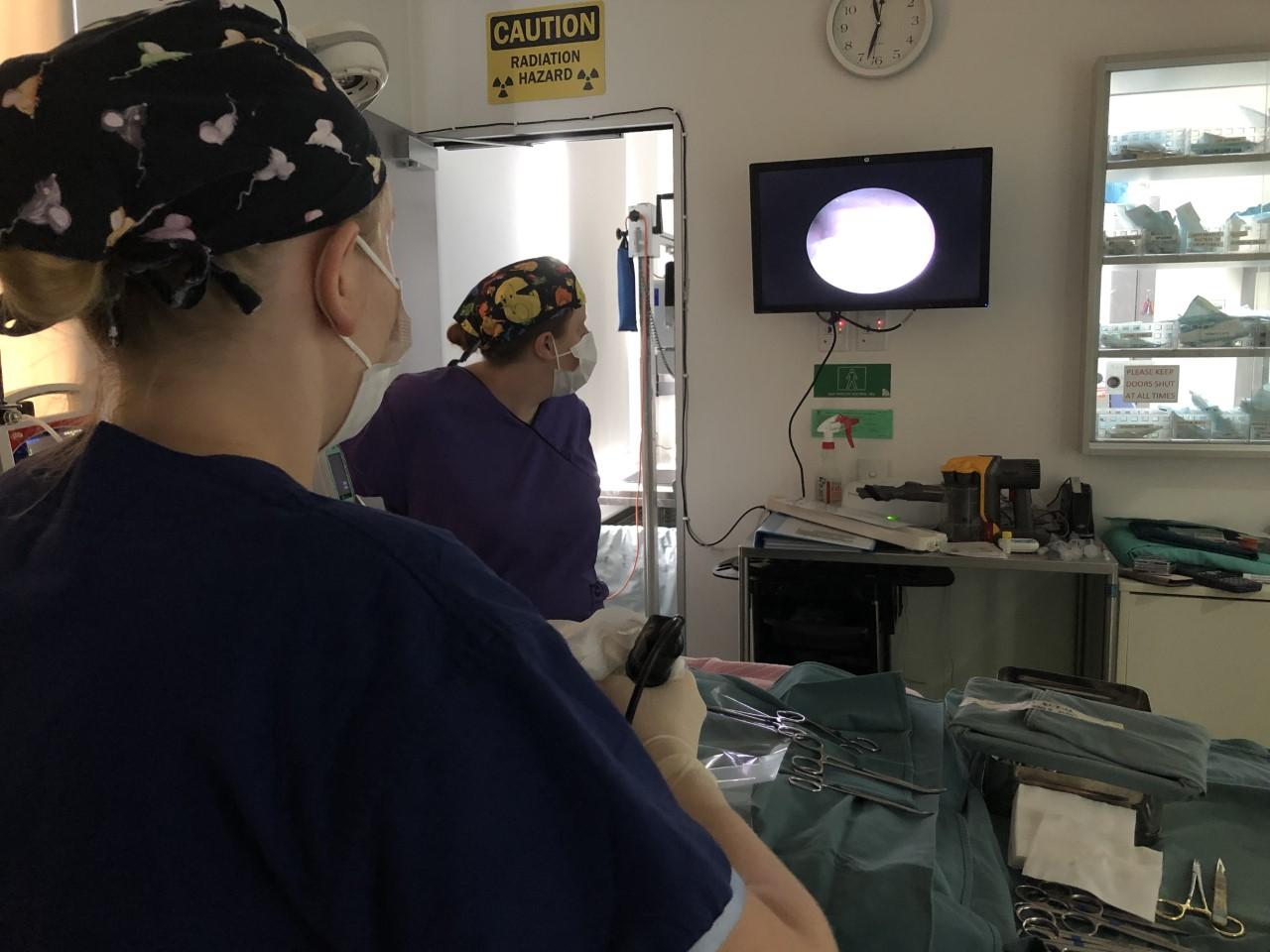
Here you can see the camera image is displayed on the monitor on the wall for easy visibility
For more information about our avian and exotic diagnostic services, just get in touch with our team through our Contact page.
To stay up to date with the latest from BBEVS, follow us on Facebook or check out our Latest News page.

